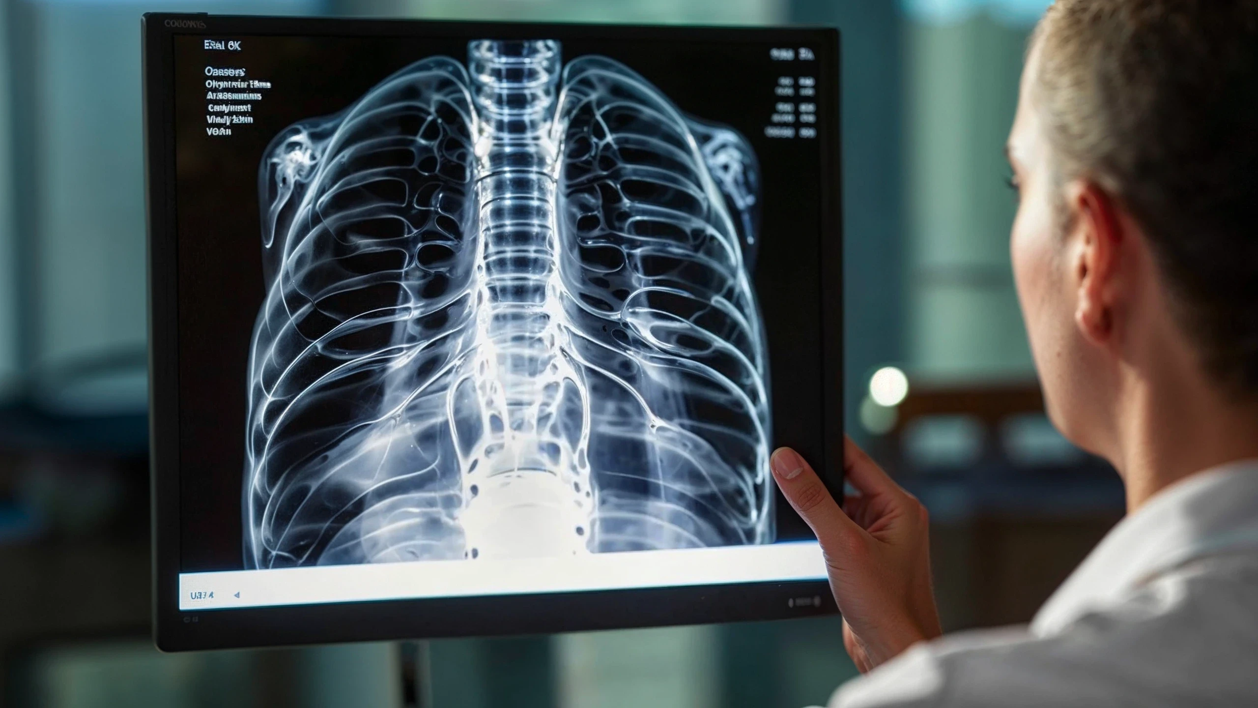
doctor examining an X-ray

Solutions

Central Imaging

Banook has dedicated the past 25 years to becoming a global leader in medical imaging technology and associated services. Whether you are a large pharma or biotech company, a CRO, or an academic institution, we can help you with your central imaging needs.

0+

0+

studies

0+

0+

studies

0+

0+

studies

0k

0k

patients

0k

0k

patients

0k

0k

patients

0+

0+

sites

0+

0+

sites

0+

0+

sites

0million

0million

images

0million

0million

images

0million

0million

images

Therapeutic areas & indications

Therapeutic areas & indications

Therapeutic areas & indications

Oncology

Cardiology

Neurology

Nephrology

Metabolic Disease

Orthopaedics

Lymphoma

Prostate cancer

Pancreatic cancer

Hepatocellular Carcinoma

Breast Cancer

Colorectal cancer

Gastric cancer

Glioblastoma

Head and Neck cancer

Melanoma

Multiple Myeloma

Non-small and small cell lung cancer

Ovarian cancer

Renal cell carcinoma

Workflow

Workflow

Workflow

Key components of a central imaging workflow

Our team provides expertise on your imaging protocol design and tailors the reading scheme to your specific imaging endpoints. We will work with you on the following key documents:

Imaging Manual: Outlines the processes to follow for site qualification, optimal image acquisition by sites, and quality control of the imaging data.

Imaging Manual: Outlines the processes to follow for site qualification, optimal image acquisition by sites, and quality control of the imaging data.

Imaging Manual: Outlines the processes to follow for site qualification, optimal image acquisition by sites, and quality control of the imaging data.

Imaging Review Charter: details the independent central read procedure of study images, including the management of central readers, the reading methodology and its rationale, and the reading schedule.

Imaging Review Charter: details the independent central read procedure of study images, including the management of central readers, the reading methodology and its rationale, and the reading schedule.

Imaging Review Charter: details the independent central read procedure of study images, including the management of central readers, the reading methodology and its rationale, and the reading schedule.

Dosimetry Documentation: describes dosimetry equipment calibration procedures, specific imaging acquisition requirements, and how to analyse the data for dosimetry analysis.

Dosimetry Documentation: describes dosimetry equipment calibration procedures, specific imaging acquisition requirements, and how to analyse the data for dosimetry analysis.

Dosimetry Documentation: describes dosimetry equipment calibration procedures, specific imaging acquisition requirements, and how to analyse the data for dosimetry analysis.

woman working on her computer

Expertise

Expertise

Expertise

radiologist operating the machine

Central  imaging reader group

Our global panel of 500+ board-certified experts—each with over 10 years of experience—ensures accurate, specialized readings across therapeutic areas. Experts are radiologists, oncologists, neuroradiologists, nuclear medicine physicians, dermatologists and cardiologists based all over the world.

Our global panel of 500+ board-certified experts—each with over 10 years of experience—ensures accurate, specialized readings across therapeutic areas. Experts are radiologists, oncologists, neuroradiologists, nuclear medicine physicians, dermatologists and cardiologists based all over the world.

Our global panel of 500+ board-certified experts—each with over 10 years of experience—ensures accurate, specialized readings across therapeutic areas. Experts are radiologists, oncologists, neuroradiologists, nuclear medicine physicians, dermatologists and cardiologists based all over the world.

Our scientific team selects and manages central readers to ensure low variability and minimize bias throughout the study.

Our scientific team selects and manages central readers to ensure low variability and minimize bias throughout the study.

Our scientific team selects and manages central readers to ensure low variability and minimize bias throughout the study.

We ensure reading consistency through statistical monitoring, consensus sessions, regular re-training, and phantom-based bias checks for quantitative imaging.

We ensure reading consistency through statistical monitoring, consensus sessions, regular re-training, and phantom-based bias checks for quantitative imaging.

We ensure reading consistency through statistical monitoring, consensus sessions, regular re-training, and phantom-based bias checks for quantitative imaging.

Reading criterias

Reading criterias

Reading criterias

Accurate imaging starts with the right reading criteria

We have supported over 350 imaging studies, many in oncology—including pivotal trials—giving us extensive expertise in the following reading criteria:

RECIST 1.1

RECIST 1.1

RECIST 1.1

Cheson

Cheson

Cheson

iRECIST

iRECIST

iRECIST

Lugano

Lugano

Lugano

mRECIST

mRECIST

mRECIST

LYRIC

LYRIC

LYRIC

PERCIST

PERCIST

PERCIST

IMWG

IMWG

IMWG

irRECIST

irRECIST

irRECIST

CHOI

CHOI

CHOI

PCWG3

PCWG3

PCWG3

Macdonald

Macdonald

Macdonald

RANO, RANO-BM

RANO, RANO-BM

RANO, RANO-BM

INRC

INRC

INRC

WHO

WHO

WHO

Tumor Volumetrics

Tumor Volumetrics

Tumor Volumetrics

We leverage our expertise to guide sponsors in selecting and implementing the most effective criteria to ensure high-quality, consistent data that supports confident decision-making.

doctor at a patient appointment

Central Imaging Modalities

Central Imaging Modalities

Central Imaging Modalities

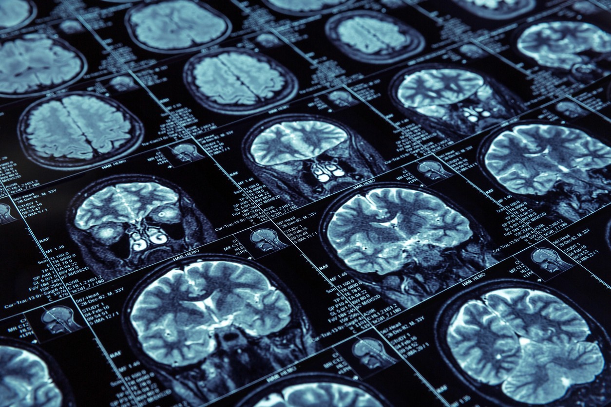
brain x-ray images
brain x-ray images
brain x-ray images

Choosing the right image modality is crucial for achieving trial objectives

We can help you with trials involving all imaging modalities including:

CT

CT

CT

MRI

MRI

MRI

PET

PET

PET

SPECT

SPECT

SPECT

X-RAY

X-RAY

X-RAY

Colonoscopy

Colonoscopy

Colonoscopy

Ultrasound

Ultrasound

Ultrasound

Angiography

Angiography

Angiography

CMRI

CMRI

CMRI

DXA

DXA

DXA

We collaborate closely with clinical teams during the protocol definition phase to ensure that all imaging challenges are addressed.

Central imaging in clinical trials

Central imaging in clinical trials

Central imaging in clinical trials

Why is central imaging important in clinical trials?

Central imaging is crucial in clinical trials as it ensures consistency in image acquisition and interpretation across multiple sites. By standardizing processes, it reduces variability, minimizes reader bias, and enhances data quality.

This approach improves the reliability of imaging endpoints, making them more robust for statistical analysis. Moreover, central imaging plays a key role in supporting regulatory submissions by ensuring the integrity, accuracy, and traceability of data.

With a centralized system, you can confidently present consistent, high-quality imaging results that meet the stringent requirements of regulatory authorities.
hands working on a tablet
Talk to an expert

Our scientific team will support you from study protocol to completion.

Contact us

Talk to an expert

Our scientific team will support you from study protocol to completion.

Contact us

Talk to an expert

Our scientific team will support you from study protocol to completion.

Contact us

FAQ

FAQ

FAQ

Frequently asked questions about Central Imaging

What types of clinical trials require central imaging?

What types of clinical trials require central imaging?

What types of clinical trials require central imaging?

How is image quality controlled centrally?

How is image quality controlled centrally?

How is image quality controlled centrally?

How long does it take to process centrally read images?

How long does it take to process centrally read images?

How long does it take to process centrally read images?